Rotator cuff tendinopathy ranks among the most common shoulder overuse injuries. Repetitive overhead movement, poor shoulder mechanics, and sudden spikes in workload frequently trigger tendon overload.
Approximately 30% of adults experience rotator cuff-related shoulder pain at some point.
Recovery requires patience, structure, and consistency rather than quick fixes.
Most individuals achieve meaningful improvement through conservative exercise-based rehabilitation without surgical intervention.
Long-term outcomes improve when rehabilitation respects tissue healing timelines and movement quality.
What Is Rotator Cuff Tendinopathy

Rotator cuff tendinopathy develops when shoulder tendons lose their ability to tolerate normal mechanical stress. Gradual overload rather than a single traumatic event drives most cases.
Repeated strain alters tendon structure, reduces resilience, and compromises force transfer across the shoulder joint.
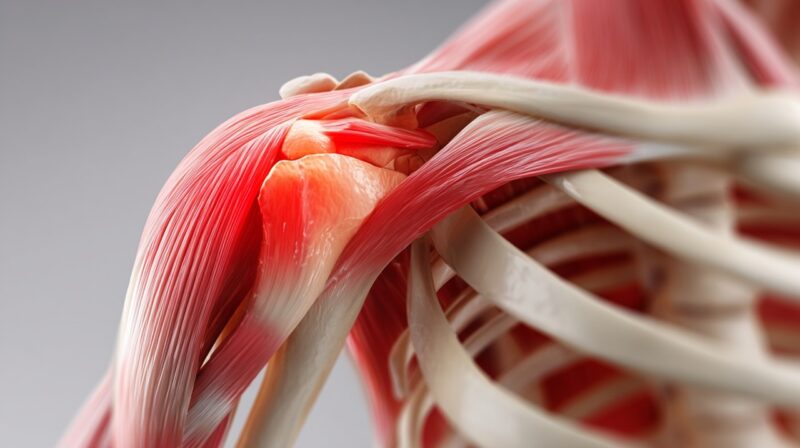
Rotator cuff anatomy consists of four primary muscles working together to stabilize and mobilize the shoulder.
Key muscular roles include:
- The supraspinatus initiates shoulder abduction and assists in humeral head compression
- Infraspinatus controls external rotation and posterior stability
- Teres minor supporting external rotation during elevated arm positions
- Subscapularis produces internal rotation and anterior stabilization
Coordinated activation maintains proper humeral head alignment within the glenoid during dynamic movement and resisted tasks.
Tendinopathy describes structural tendon degeneration marked by disorganized collagen, reduced tensile strength, and diminished load tolerance rather than active inflammation.
Tendonitis represents an inflammatory response and typically appears only during early or acute overload phases.
Partial or full-thickness tears involve the disruption of tendon fibers and significantly prolong recovery due to impaired mechanical continuity.
Multiple contributors interact to exceed tendon recovery capacity. Repetitive microtrauma accumulates when workload increases outpace tissue adaptation.
Biomechanical inefficiencies amplify compressive and tensile stress on rotator cuff tendons.
Common contributing factors include:
- Altered glenohumeral mechanics and poor scapular control
- Limited thoracic spine mobility is restricting overhead mechanics
- Age-related degeneration increases after forty years
- Metabolic influences such as diabetes and elevated cholesterol
- Reduced tendon blood supply associated with smoking
- Occupational or athletic demands involving overhead or throwing tasks, such as those found in archery lessons
- Forward head posture and rounded shoulders narrow the subacromial space
Common Signs and Symptoms
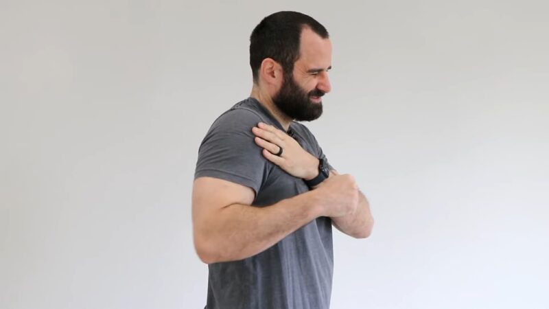
Symptom presentation varies but follows recognizable patterns tied to tendon load sensitivity. Pain typically develops gradually and intensifies during tasks requiring elevation or sustained arm use.
Most individuals report hallmark features that indicate rotator cuff involvement:
- Pain during overhead activity within a painful arc between sixty and one hundred twenty degrees of abduction
- Night pain or discomfort while lying on an affected shoulder
- Weakness during lifting, reaching, or rotational movements
Functional changes extend beyond pain alone. Clicking, popping, or crackling sensations may accompany movement due to altered tendon glide or joint mechanics.
Shoulder endurance often declines, leading to faster fatigue during daily or athletic tasks. Local tenderness develops over involved tendons, particularly after activity.
Symptom flares frequently follow abrupt increases in training volume, workload intensity, or frequency when tissues lack adequate adaptation time.
Diagnosis and Imaging
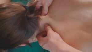
Accurate diagnosis relies on combining clinical assessment with appropriate imaging when needed. Physical evaluation focuses on movement quality, strength symmetry, and symptom reproduction under controlled load.
Clinical testing serves specific diagnostic purposes:
- Drop arm testing screens for significant supraspinatus dysfunction
- Empty can testing evaluates supraspinatus strength and pain response
- Hawkins Kennedy testing assesses subacromial impingement sensitivity
- Manual resistance testing identifies strength deficits across planes of motion
Imaging assists when structural information alters management decisions. Ultrasound allows real-time assessment of tendon thickness, integrity, and dynamic movement.
Magnetic resonance imaging identifies tear size, location, and tendon quality with high sensitivity.
X-rays help rule out bony contributors such as osteophytes or degenerative joint changes that may worsen impingement.
Accurate diagnosis supports appropriate load management exercise selection and recovery pacing. Identification of biomechanical postural and systemic contributors reduces recurrence risk and improves long-term outcomes.
Phases of Healing and Expected Timelines
Tendon healing follows predictable biological stages that influence rehabilitation progression. Respecting these phases prevents overload during vulnerable periods and supports durable recovery.
Early biological responses involve distinct tissue changes:
- Inflammation during the first seven days is characterized by pain, swelling, and immune activity
- Early healing between one and six weeks, as collagen synthesis begins
- Remodeling between six and twelve weeks with improving tissue organization
- Maturation spanning three to twelve months or longer, with gradual restoration of tendon capacity
Recovery duration depends on severity, tissue quality, and adherence to rehabilitation principles.
Minor presentations often resolve within four to six weeks using structured exercise.
Moderate cases usually require three to six months for full functional recovery.
Surgical cases demand six to twelve months or longer, depending on tear size, tissue quality, and rehabilitation consistency.
Non Surgical Rehabilitation Plan
Exercise based rehabilitation serves as primary treatment even with partial tendon tears.
Progressive mechanical loading stimulates collagen alignment restores strength and improves tendon tolerance.
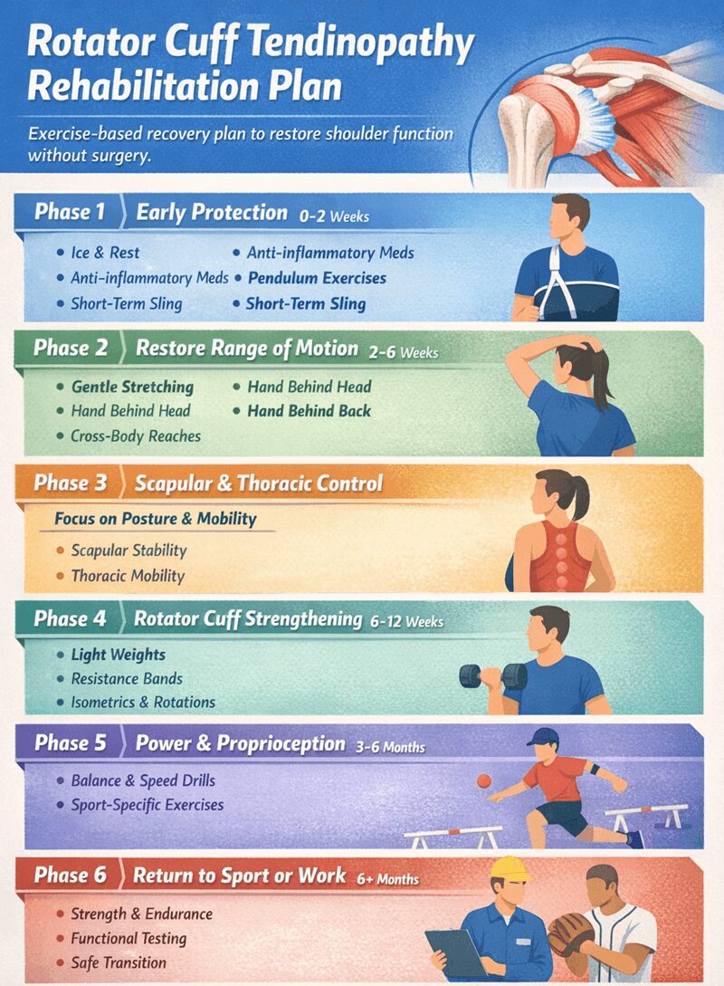
Phase One Early Protection Zero to Two Weeks
Initial care focuses on symptom control and protection without complete inactivity. Relative rest reduces excessive load while maintaining joint health.
Early strategies include:
- Ice application for twenty to thirty minutes two to four times daily
- Anti-inflammatory medication during acute pain phases when appropriate
- Gentle pendulum exercises supporting joint nutrition and mobility
- Short term sling use limited to comfort needs
Phase Two Restore Range of Motion Two to Six Weeks
Mobility restoration prevents stiffness and supports later strengthening. Active assisted movement gradually replaces passive strategies as pain allows.
Priority movements include hand behind head, hand behind back, and cross-body reaching. Soft tissue techniques reduce adhesions and promote tissue glide.
Pain-free range determines progression speed.
Phase Three Scapular Control and Thoracic Mechanics
Efficient shoulder movement depends on coordinated scapular and thoracic motion.
Scapulohumeral rhythm retraining improves force distribution during elevation.
Thoracic spine mobility and cervical posture receive targeted attention. Postural correction reduces subacromial compression and improves load sharing.
Phase Four Rotator Cuff Strengthening
Strength development begins with low load activation and advances toward controlled resistance. Loading strategies progress deliberately to avoid reactive pain.
Effective exercises include:
- Isometric contractions during early strengthening
- Side-lying dumbbell external rotation for posterior cuff engagement
- Side-lying eccentric adduction challenging controlled tendon loading
- Resistance band external rotation building endurance
Humeral head centralization remains a priority throughout all exercises.
Phase Five Proprioception Power and Speed Three to Six Months or Longer
Advanced rehabilitation introduces velocity coordination and reactive control.
Functional loading prepares tissues for real world demands.
Sport or job specific drills restore movement efficiency. Power speed and joint position awareness reduce reinjury risk during unpredictable tasks.
Phase Six Return to Sport or Work
Return progression respects fatigue thresholds and workload tolerance. Movement mechanics, endurance, and strength guide readiness decisions. Performance testing confirms capacity. Collaboration with coaches or employers supports a safe transition.
Surgical Options When Conservative Care Fails
Surgical intervention becomes necessary for a minority of cases.
Approximately 20% require operative management due to structural damage or persistent dysfunction.
Surgical consideration arises with:
- Large or full-thickness tendon tears
- Symptoms persisting longer than six months despite rehabilitation
- Significant strength loss or functional limitation
- Acute traumatic tears in younger athletic populations
Post-surgical rehabilitation follows a staged progression. The protection phase spans zero to six weeks with sling use and passive motion only. The early motion phase occurs between six and twelve weeks, introducing assisted then active movement.
The strengthening phase develops between three and six months using progressive resistance. Return to sport or work typically occurs between six and twelve months with functional training and clearance.
The highest retear risk occurs between three and five months post-surgery when pain improves faster than tendon strength. Careful load management during this window remains critical.
Summary
Rotator cuff tendinopathy responds well to structured rehabilitation aligned with tissue biology.
Successful recovery depends on load progression, movement quality, and consistency.
Injection-based shortcuts offer limited long-term benefit unless medically indicated.
Proper rehabilitation restores pain-free function, reduces recurrence risk and supports durable shoulder health for athletes and workers alike.